Our Services
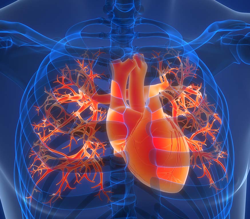
Cardiac Imaging

Coronary Calcium Scan
Coronary artery disease occurs when plaque builds up and narrows the arteries that supply blood to your heart (atherosclerosis) and is the leading cause of heart attacks.
A Coronary Calcium Scan measures the amount of plaque or calcium in the walls of your coronary arteries. It provides a calcium score, which your cardiologist will use alongside other health information to determine your future risk of coronary artery disease or heart attack before you develop any symptoms.
You should consider having a coronary calcium scan if:
- You are a man over 45 years of age
- You are a woman over 55 years of age
- You have at least one of the following risk factors:
- Family history of heart disease
- High cholesterol
- High blood pressure
- Diabetes
- Smoker
The procedure takes about 5 minutes, involves no injections and does not require you to fast beforehand.
Coronary Computed Tomography (CTA)
Coronary CTA provides pictures of the coronary arteries, which supply blood to the heart muscle, in a non-invasive manner. It enables your cardiologist to look for narrowings, some of which cannot be detected by the more traditional exercise testing, but which cause around three quarters of all heart attacks. Detecting these narrowings early is important as medications can be prescribed to prevent an impending heart attack. Based on the results from the CTA, your surgeon will order further tests to decide on any course of treatment.
We use the latest generation, state-of-the-art Toshiba 64-Slice Coronary CTA scanner, which can simultaneously acquire 64 image slices of the heart in one minute, resulting in highly detailed images of the coronary arteries and the heart.

Cardiac MRI (Magnetic Resonance Imaging)
Cardiac MRI (Magnetic Resonance Imaging) is a non-invasive imaging technique used to assess the structure and function of the heart. It provides high-resolution images and detailed information about the heart and surrounding blood vessels.
Assess Cardiac Anatomy
- Heart Chambers and Valves: Cardiac MRI provides a detailed view of the heart’s chambers, evaluates the size, shape, and function of these chambers, as well as the heart valves.
- Wall Thickness: MRI can measure the thickness of the heart muscle (myocardium). Abnormal thickening or thinning of the heart wall may indicate diseases like hypertrophic cardiomyopathy or dilated cardiomyopathy.
Assess Cardiac Function
- Ejection Fraction (EF): One of the most important metrics for evaluating how well the heart pumps blood. Cardiac MRI gives highly accurate measurements of the left ventricular ejection fraction (LVEF), which is the percentage of blood the left ventricle pumps out with each heartbeat.
- Cardiac Output: It can calculate the amount of blood the heart pumps per minute, which is an important indicator of heart function.
Evaluate Specific Heart Conditions
- Ischemic Heart Disease: Cardiac MRI can evaluate the extent of damage caused by a heart attack (myocardial infarction), including the location of infarcted (dead) tissue, scar tissue, and viable myocardium (healthy heart muscle).
- Cardiomyopathies: MRI can help diagnose various forms of cardiomyopathy, such as:
- Dilated Cardiomyopathy: where the heart chambers become enlarged and weakened.
- Hypertrophic Cardiomyopathy: where the heart muscle becomes abnormally thickened.
- Restrictive Cardiomyopathy: where the walls of the heart become stiff and less able to expand and contract.
- Inflammatory Conditions: MRI is highly effective at detecting inflammation in the heart, such as in cases of myocarditis (inflammation of the heart muscle) or sarcoidosis.
Evaluate Pericardium
- Pericardial Disease: MRI can provide detailed images of the pericardium (the membrane surrounding the heart), helping detect conditions like pericardial effusion (fluid around the heart), pericarditis, or constrictive pericarditis.
Cardiac Tumors and Masses
- Detection of Tumors: Cardiac MRI is highly effective in identifying cardiac tumors, such as benign or malignant growths, including myxomas, which are the most common type of primary heart tumor.
Congenital Heart Disease
- Congenital Defects: Cardiac MRI can be used to assess congenital heart defects (conditions present from birth), such as septal defects (holes in the heart), abnormal connections between vessels, or other structural abnormalities.
In Summary
Cardiac MRI is invaluable for diagnosing a wide range of heart conditions. Its ability to offer non-invasive, high-resolution images with no ionizing radiation makes it an excellent choice for heart health evaluation and ongoing monitoring.
Unsure if you need Cardiac Imaging?
Our cardiologists can help determine the cause of your symptoms and recommend the right tests and treatment.
Risk Factors of Heart Disease
- Family history of heart disease
- High cholesterol
- High blood pressure
- Diabetes
- Smoker
Schedule A Heart Health Check Today
Your heart works hard for you—make sure it’s in good shape. A quick check-up today could help prevent serious problems tomorrow.
-
Monday to Friday
8.00 am to 5:00 pm
-
Saturday
8:30 am to 12:30 pm
-
Sunday
Closed
-
Monday to Friday
8.00 am to 5:00 pm
-
Saturday
8:30 am to 12:30 pm
-
Sunday
Closed
-
Monday to Friday
8.00 am to 5:00 pm
-
Saturday & Sunday
Closed
-
Monday to Friday
8.00 am to 5:00 pm
-
Saturday
8:30 am to 12:30 pm
-
Sunday
Closed